Sunday, December 18, 2011

Frog Dissection Lab


Photo From: http://dnr.wi.gov/eek/critter/amphibian/leopard.htm

Over a course of two days, our class completed a frog dissection lab. The purpose of this lab was to have a better understanding of the organs and parts of the frog, the functions of those organs and parts, and how they connect with us. On the first day, we focused on cutting open the frog, taking out the fat bodies, and taking out the eggs and oviducts if the frog was female. I was surprised how strong the skin of the frog was. I had expected it to cut open easily, but it actually took our group a few tries to finally cut it. After the skin, was a layer of muscle, which actually seemed easier to cut, but that may have been because our group was used to cutting by then. The first thing I noticed after the frog was cut open, was the eggs. There were black spherical eggs everywhere in the frog. This told us that we had a female. Taking out the eggs required patience and perserverance. As soon as you thought you had finished taking out all of the eggs, you'd find more. It almost seemed never ending. I envied the groups that had male frogs and didn't need to clean out all the eggs. Eventually, we focused on clearing out the fat bodies and oviducts. There were still a few eggs scattered around, but we still had visibility of the organs and parts. That was the end of day one.


Picture From: http://www.realfishbaitcompany.com/products/

On day two, we focused on identifying the parts of the frog, which we had named Kermit, and the functions of those parts. For me, the pancreas and kidney were the hardest to tell apart. They seem so similar, although they have different functions. What surprised me the most was how small the lung seemed to be. After all, the lungs hold oxygen. It's hard to imagine it expanding into anything large enough to hold oxygen for a frog. In addition, I thought the gall bladder would be bigger. The liver, which produces bile, is the largest structure in the body cavity, but the gall bladder, which stores the bile, is about the size of a pea. This dissection also caused me to realize that the parts of the frog are more complex than the worms, and similar to ours (such as the digestive system). Overall, this frog dissection lab was very interesting, as well as surprising, and gave me a better understanding of the frog parts and functions, and how they connect with us.


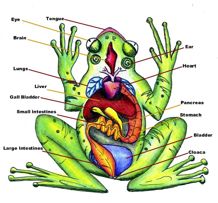
Right Image from: http://www.biologyjunction.com/frog_dissection.htm


For more information on frogs, and frog dissection visit:

http://www.biologyjunction.com/frog_dissection.htm

http://www.e-tutor.com/et3/lessons/view/52133/print

http://www.ecokids.ca/pub/eco_info/topics/frogs/quiz/quiz_3.cfm

http://www.biologycorner.com/bio2/notes-frog.html


Tuesday, November 8, 2011

Digestion Lab

The past two days, we have conducted a lab using, hydrochloric acid, pepsin, boiled egg whites, and water. In four test tubes, labeled A, B, C, and D, we put in three pieces of egg white. In test tube A, we put 10 mL of pepsin. To test tube B, we added 5 mL of water and 5 mL of pepsin. Next, in test tube C, we added 10 mL of hydrochloric acid. Finally, 5 mL of pepsin and 5 mL of hydrochloric acid were added to test tube D. We then had to observe whether a change happened immediately. In test tubes A and C, there was no immediate reaction, but in test tube B, it turned foggy. In addition, in test tube D, it looked as if the egg was slowly melting or turning mushy, Next, we took a straw and dipped it into each test tube then touched the straw to a piece of blue lithmus paper to see if the paper changed color. In test tubes C and D, the paper turned pink. The other test tubes stayed the same.
The next day, we observed our test tubes to see if they had changed. Test tubes A and C, it looked the same. The egg whites in test tube be looked more delicate so if you were to squish them, they'd mush together. In test tube D, the mixture was foggy and it looked as if the egg whites had dissolved and piled together in a mush at the bottom. Also, we had to dip the straws in the mixture again and place it onto the lithmus paper. Test tubes A and B didn't change the color of the lithmus paper, but test tubes C and D made the paper pink again. This lab showed that our stomach was like test tube D. It was full of enzymes (pepsin) and hydrochloric acid working together to digest the food that we eat.

Thursday, October 20, 2011

Chicken Wing Anatomy Lab

Today, Mrs. Rousseau dissected a chicken wing to show the different tissues and organs and how they work. The skin that protects the chicken's tissues and organs is epithelial tissue. Also, the muscle in the chicken wing was the actual tissue that moved the chicken wing by tightening and loosening. Other parts of the chicken wing that we saw were the fat and bone, which are both connective tissues. The only type of tissue we didn't see was the nerve tissue.

Tuesday, October 11, 2011

Diffusion Lab

Today, we had to do a diffusion lab to observe the diffusion of a substance across a semi permeable membrane. We mixed water and cornstarch in a baggie and submerged it in a beaker filled with water and an iodine solution. Then we waited 15 minutes. We added iodine as an indicator for the starch (a substance that changes color in the presence of the substance it indicates). After 15 minutes, we checked on our cornstarch baggies. The lab didn't go as expected. The iodine mixture didn't really enter the baggie and change the color of the water and corn starch mixture. I think this was because the plastic baggie was made a lot stronger so the iodine solution couldn't enter it.